Advanced Technology Aids in Proper Diagnosis
“Pain related to the temporomandibular joint (TMJ) is common in the general population,” states an article published by the National Institutes of Health’s National Library of Medicine. Yet only about three to seven percent of the patients with associated pain seek medical attention.
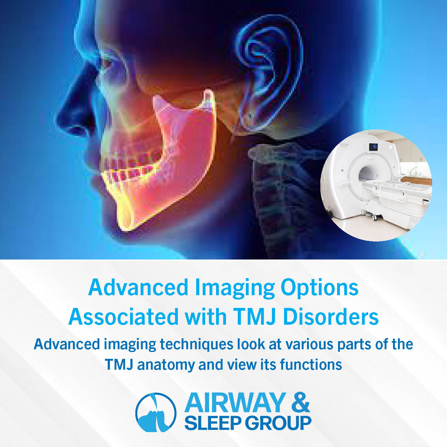
Advancements in imaging of the temporomandibular joint are helping dental and health professionals properly diagnose TMJ disorders and better understand the biomechanics of the TMJ. The modalities include non-invasive imaging methods such as radiographs, ultrasound, CT, and MRI, all the way to invasive imaging like arthrography.
Radiographs
Conventional radiographs have a limited role. They evaluate only the bony element of the TMJ.
Ultrasound
Ultrasounds are easily performed and are a less-expensive option when looking for the presence of joint effusion, or evaluable cartilage or disk displacement with both open and closed mouth imaging. It can be used for image-guided injections.
CT
CT, or computed tomography, helps evaluable both the bony elements of the TMJ and the adjacent soft tissues. It’s ideal for evaluating fractures, degenerative changes, erosions, infection, tumor invasion, and congenital anomalies.
MRI
MRI, or magnetic resonance imaging, provides high resolution and tissue contrast to aid in the clinical evaluation of the TMJ. It allows a detailed evaluation of the anatomy and the jaw biomechanics in open and closed mouth imaging.
TMJ disorders or dysfunctions are the most common clinical conditions for imaging referrals, but pathologies specific to the bone and joints are also common. Different imaging modalities are used to view the temporomandibular joint, each with its inherent strengths and weaknesses. Magnetic resonance imaging, or MRI, is the most widely used and the diagnostic technique of choice due to its superior contrast resolution and its ability to acquire dynamic imaging for demonstration of the functionality of the joint.
Arthrography
Arthrography is an imaging test that shows a detailed x-ray image of the inside of a joint structure and function and is used in the evaluation of joints to locate hard-to-find problems. It’s conducted by the insertion of a needle into the joint and the injection of a contrast medium into the joint cavity.
What Imaging Shows
Jaw movement involves a high level of interaction and coordination between bilateral mandibular condyles, disks, muscles, and ligaments of the joints. A health professional uses advanced imaging techniques to look at various parts of the TMJ anatomy and to also view its functions.
Items reviewed include the:
- Articular disk (the stress absorber)
- Muscles
- Adductors/jaw-closing muscles
- Abductors/jaw-opening muscles
- Joint ligaments
What are TMJ and TMJ disorders?
The temporomandibular joint, or TMJ, is a joint in your facial skeleton that attaches the jawbone to the skull. There’s one joint on each side of the jaw that acts like a sliding hinge that allows movement of the jaw both backward and forward, as well as a gliding motion.
During gestation, the TMJ is one of the last joints to appear in utero, often emerging in the 8th week. It’s also “considerably underdeveloped at birth in comparison to other diarthrodial joints, making it susceptible to perinatal and postnatal insults,” the article states. It continues to develop into the early childhood years as the jaw is used for sucking motions and then chewing.
A TMJ disorder, which is sometimes mistakenly called “TMJ,” is created by a malfunction of that joint, and is evident by pain in the joint and/or the muscles that control the jaw movement. The cause, however, is difficult to determine. The pain may be caused by an injury, clenching or grinding of teeth, genetic factors, or arthritis, for example.
In most cases, the pain and discomfort can be remediated with nonsurgical treatment or self-care. Surgery is the last choice when other treatments are not effective.
TMJ Disorder Signs & Symptoms
The symptoms of a TMJ disorder can include:
- Jaw pain or tenderness
- TMJ joint pain
- Pain in or around the ear
- Difficulty chewing, or painful chewing
- Joint locking
- Clicking or grinding when opening or closing the mouth or chewing
Diagnose a TMJ Disorder and Receive Treatment at Sleep & Airway Group
If you’d like to learn more about TMJ pain or suspect you have a TMJ disorder, talk to the professionals at Sleep & Airway Group in Reston, VA. Dr. Liliana Calkins, DDS is a board-certified orthodontist and is trained in craniofacial development and dentofacial orthopedics. You can also fill out our online contact form and request more information.
–information excerpted from “Imaging of the temporomandibular joint: An update,” published by the NIH’s National Library of Medicine.