Listen to our podcasts here.
In this Airway and Sleep Group Podcast, Dr. Liliana Calkins interviews Dr. Francisco Eraso. Dr. Francisco Eraso is a specialist in orthodontics and oral and maxillofacial radiology. A member of the American Association of Orthodontics, American Academy of Oral Maxillofacial Radiology, and the International Association of Dental Maxillofacial Radiology. He is a practitioner in Indianapolis and one of the founders of Beam Readers Diagnostic Services.
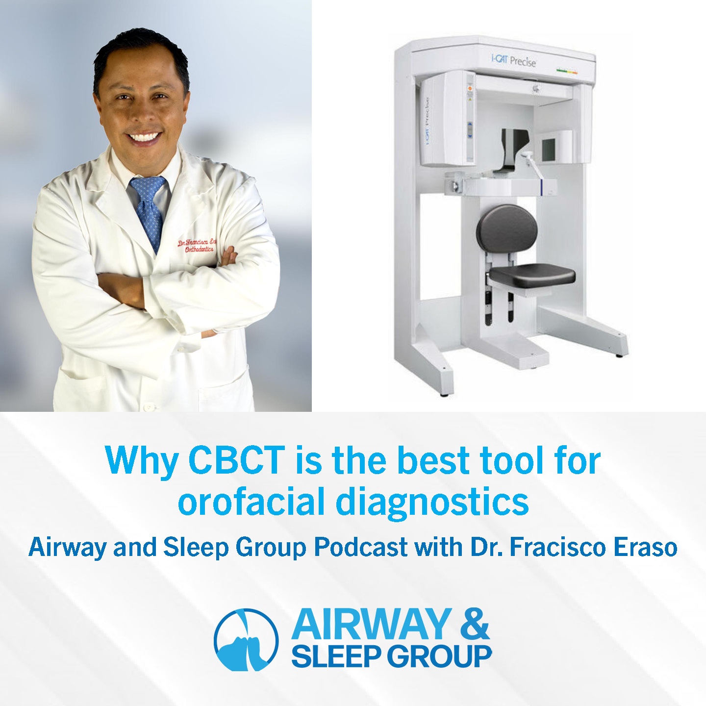
Why do you consider CBCT an essential diagnostic tool for imaging the anatomy of the craniofacial complex within dentistry or the specialty of orthodontics?
Knowing anatomy is power. If you know your anatomy, you can derive different solutions. Anatomy points you in the right direction for TMJ, orthodontics, and airway. To be successful in orthodontics, you have to know your anatomy and the impact it has on the orofacial complex.
Teeth are not only to be straightened. An orthodontist who thinks they can do everything with aligners is wrong. The knowledge learned about a patient’s basic oral anatomy is essential in creating a diagnostic plan.
A lot of people do aligners based on crowns by taking scans and panoramics, however, in the panoramic you don’t see the boundaries of the mandibular bone and you pretend that you’re going to move teeth. Sometimes you’re lucky and you have some movements, however, if you don’t know where the root is in the bone it’s very difficult to be efficient in the movement. The knowledge of where the roots are positioned in the mandibular bone is essential.
Why do you need 3D imaging for diagnostics?
CBCT is an essential diagnostic tool for imaging the anatomy of both the mandibular bone and the maxillofacial complex. We’re not just teeth and bone, but we’re surrounded by amazing structures like the airway, TMJ, and maxillary sinuses. The only tool that can show you the 3D structure is CBCT.
You also learn about the floor of the sinus, which is within a close relationship with the roots of the molars. Sometimes it’s difficult to move the molars in the mesial direction or the distal direction because of the relationship of the roots with the cortical alignment on the floor of the maxillary sinus.
If you don’t have a 3-dimension knowledge, image, or interpretation of where the roots are, you’re going to be blind in creating your treatment plan. You’re going to move teeth in the computer, the crowns are going to move fantastically in the software but in reality, the treatment is going to be very inefficient.
So if you don’t take into account 3D anatomy, like with Invisalign, or any kind of aligner software, sometimes you have to do an infinite number of refinements to orthodontic aligner treatment. Some people think that’s fine, but in actuality, it’s orthodontists that aren’t doing a good job because they didn’t study anatomy. That’s why using a diagnostic tool to uncover the underlying anatomy is essential in developing an effective orthodontic plan that achieves success.
What is the trend that you see in the adoption of CBCT by dental care physicians and the implementation of radiology reports?
Over the years we create habits and the habit of every dentist is to take a panoramic x-ray, also known as bite-wings. Most dental offices are equipped with these two diagnostic tools. However, these tools won’t give you anything. The only thing you can get from a panoramic is to count teeth. It won’t give you a good localization of impact teeth, or an excellent view of the temporomandibular joint, the maxillary sinus, or the airway.
Today, we don’t have an excuse not to have a CBCT, because CBCT’s are used everywhere. Getting a CBCT is like knowing the truth. The radiologist will give the knowledge to you, you just cannot fear knowing the truth. You want to have the best tools for diagnosis.
How can you do the diagnosis and treatment plan without the best tools?
Do you have a specific case that revealed an unexpected finding?
We read 100,000 cases with 50 radiologists. I read about 8,000-10,000 cases per year. One of the classic findings is a case of hyper-density that surrounds the roots, which is shown as white on the CBCT. If you take radiographs you won’t see the extent of that opacity or hyper-density. This condition is called a dense bone island, enostosis, idiopathic osteosclerosis. For you to move a tooth through that bone, it will take time. Or you have to conclude that that tooth isn’t going to move and build your diagnostic plan around it.
It’s good to take a high-quality image at the beginning than a reactive image in the middle of the treatment. You don’t want to be surprised by the anatomic structures after the treatment plan has begun, you want to have the knowledge and the truth of the anatomy before you begin to make a good diagnostic plan.
What would you say to patients who are hesitant to get a CBCT for orthodontic diagnostics?
For patients who are hesitant to obtain CBCT imaging due to radiation exposure, you can expect about the same amount of radiation emitted from a panoramic x-ray. In an imaging portfolio, you extract information about the airways, TMJ, soft palate, nasal cavity, maxillary sinuses temporomandibular joint, rule out pathology in the bone and impacted teeth. Additional findings include the classification of the arteries and tonsils. It can also predict the appliance’s effect in the mechanics at the end of the treatment — the effect of the airway spaces, the improvement of the facial soft tissue, and more.
In short, specific information leads to making good decisions, which far outweighs the risk of a radiation dose. A CBCT has a near equivalent amount of radiation transmitted from a panoramic x-ray, and the truth and information gained from a CBCT reduce your chances of creating a diagnostic plan that’s not feasible and reduces the need for multiple orthodontic refinements.
Visit Dr. Calkins Today
Airway and Sleep Group has state-of-the-art technologies that give us the most accurate diagnosis available and help us get the most consistent, beautiful, smiles possible! Schedule a consultation for your orthodontic and airway diagnosis by visiting www.airwayandsleepgroup.com.
You can also give our Reston office a call at (571) 244-7329 or fill out our online contact form. We look forward to speaking with you.